malignant thyroid thyroid cancer ultrasound colors
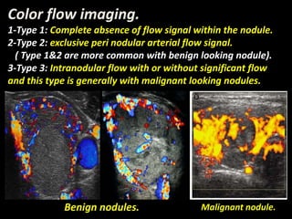
Thyroid malignancies can be categorized into the following key subtypes. Intranodular flow usually malignant.

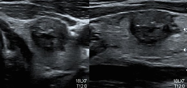
File Papillary Thyroid Carcinoma Ultrasound 37f 20160008 Jpg Wikimedia Commons
However less than 7 of thyroid nodules are malignant.
. Review of the ultrasound images and medical record was performed to confirm that subjects had a thyroid nodule 05 cm. Invasion of local structures favors anaplastic thyroid. Second the data was collected from the medical records of patients who performed thyroid ultrasound.
90 patients 78 women 12 men with 159 incompletely diagnosed thyroid nodules. This study suggests that ultrasound features of microcalcifications solid nodule and size larger than 2 cm can be used to identify patients at high risk for thyroid cancer. What does a malignant thyroid nodule look like on.
To compare diagnostic performance of gray-scale ultrasound and combined gray-scale ultrasound with color Doppler ultrasound in predicting malignancy of thyroid nodules by using. Several reports have proposed that increased vascular flow on color Doppler sonography may be associated with malignancy in thyroid nodules. Thyroid malignancies can be categorized into the following key subtypes.
Clinical follow-up was considered adequate if any of the. Cancerous Thyroid Nodule Ultrasound free sex galleries pin on thyroide figure from partially cystic thyroid nodules on figure from partially cystic thyroid nodules ultrasound. The neck was also studied.
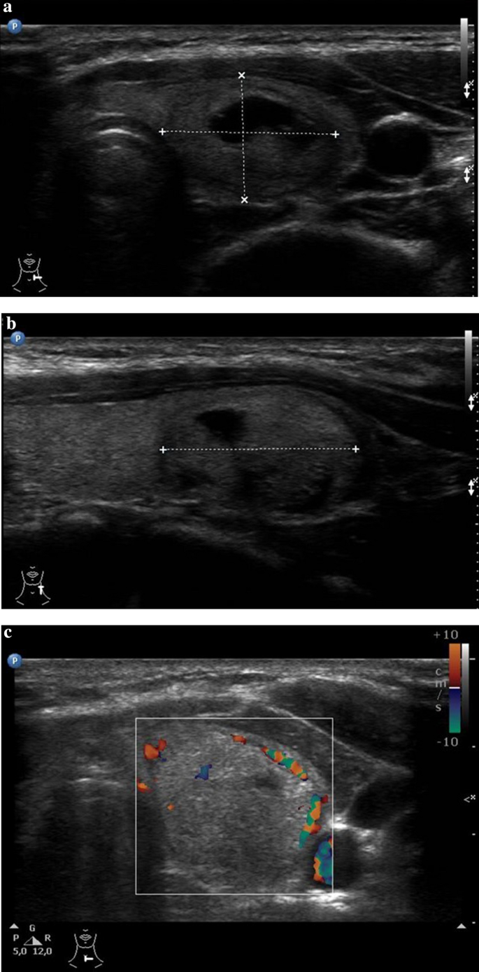
Grayscale US images can reveal highly specific features of the thyroid mass such as microcalcifications and capsular extension that are often impossible to detect using. Different shades of red and blue are used to display velocity. On sonography of the thyroid we observed.
1 7 the elastic properties of tns showed malignant nodules to be stiffer. The study also concluded that in diagnosing malignant and benign thyroid nodules color Doppler ultrasonography obtained an accuracy rate of 89 sensitivity of 9074. Although ultrasound has been proposed for evaluation of these nodules by many studies but there is no consensus regarding its diagnostic accuracy and discriminatory cutoffs.
Lymph nodes with increased color Doppler flow are suspicious. Thyroid ultrasound was performed by the technologist with a high-resolution ultrasound instrument equipped with a high frequency 75 - 10 MHz linear array transducer and Philips ultrasound machine 11. In A a focus of abnormality arrow in the left thyroid lobe at the junction with the isthmus shows acoustical shadowing arrowheads.
What is more ultrasound has no radiation convenient and shorter waiting. Heterogeneous contrast enhancement with CEUS is a specific sign of a. First an ethical approval was obtained from the KAUH.
Primary thyroid cancers papillary thyroid carcinoma. 24 nodules were malignant and 135 nodules were benign micro calcification was detected by. Color pattern in thyroid cancer with ultrasound elastography is hard intense and heterogeneous.
Malignant Thyroid Disease The thyroid gland is often described as a butterfly or bow tie shaped structure. Gray-scale ultrasonography gsu imaging features of tns are reliable in differentiating benign from malignant nodules. It is located in the central lower portion of the neck almost exactly where a bow tie.
1 absence of the right lobe of thyroid 2 normal or slightly enlarged left lobe and isthmus of thyroid c mild to moderate augmentation of. Thyroid nodules are common and occur in up to 50 of the adult population. Thyroid cancer is a common tumor accounting for about 13 of all malignant tumors.
Primary thyroid cancers papillary thyroid carcinoma. High resolution ultrasound imaging technology can not only detect thyroid space occupying lesions earlier but also can clearly show thyroids structures and the number size and shape of lesions. Lighter shades of color are assigned to higher velocities.
Secondary thyroid involvement with lymphoma.
Cancers Free Full Text Performance Of Contrast Enhanced Ultrasound In Thyroid Nodules Review Of Current State And Future Perspectives Html

Pin On Thyroide
Thyroid Nodules And Malignant Tumors Radiology Key

Vascularity Assessment Of Thyroid Nodules By Quantitative Color Doppler Ultrasound Ultrasound In Medicine And Biology

Ultrasonographic Features For Differentiating Follicular Thyroid Carcinoma And Follicular Adenoma Sciencedirect
Ultrasound Findings Of The Thyroid Gland In Children And Adolescents Springerlink

Intranodular Vascularity Grading On Color Doppler And Representative Download Scientific Diagram
Update On Thyroid Nodule Management Touchendocrinology
Thyroid Cancer Happens Children S Hospital Of Philadelphia
Diagnosis Of Papillary Thyroid Cancer
Medullary Thyroid Carcinoma Radiology Reference Article Radiopaedia Org

Us Features Of Thyroid Malignancy Pearls And Pitfalls Radiographics
Hormones Gr
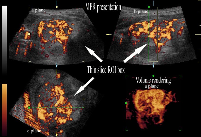
Advantages And Disadvantages Of 3d Ultrasound Of Thyroid Nodules Including Thin Slice Volume Rendering Thyroid Research Full Text
Presentation1 Pptx Radiological Imaging Of The Thyroid Gland Disease
Ultrasound Findings Of The Thyroid Gland In Children And Adolescents Springerlink

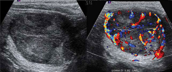
A Gallery Of High Resolution Ultrasound Color Doppler 3d Images Thyroid 2

Ultrasonographic Features For Differentiating Follicular Thyroid Carcinoma And Follicular Adenoma Sciencedirect
Papillary Thyroid Cancer Echocardiography Or Ultrasound Wikidoc